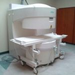
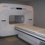
MRI can be elaborated as Magnetic Resonance Imaging. It is a scanning technique to generate a detailed image of our body. It is used in the field of diagnosis. That means, diagnose many disorders like strokes, tumours, aneurysms, spinal cord injuries, multiple sclerosis etc. It is a fast and useful innovation that helps medical professionals study body anatomy and body parts that is of interest. The scanner depends on biological parameters like longitudinal relaxation time(T1), variable image contrast, proton density & transverse relaxation time(T2) that are obtained by utilizing variable pulse sequence and changing imagining parameters. Signal intensities T1 & T2 allows superb contrast between different soft tissues.
The multiplanar capability of MRI, we can optimize the imaging plane for the particular atomic area under observation. The relationship between lesions and eloquent brain areas can be obtained precisely. Blood flow data and vascular anatomy can be obtained with the help of MR angiography and flow-sensitive pulse sequences. We can even study brain functions with the help of MRIs. MR spectroscopy can be used to study tissue metabolism and biochemistry. MRI technology is continuously evolving with advancement in medical technology.
MRI system includes the following components:
Big magnet to produce a magnetic field
Shuim coils that produce the homogeneous magnetic field
Radio frequency coil that transmits radio signals to body parts that are imaged
Receiver coil to receive reflected radio signals
Gradient coils that create spatial localisation of signals
Computer to reproduce the radio signal to the final image
A quick history of the MRI
1882 – Nikola Tesla discovered the Rotating Magnetic Field in Budapest, Hungary. This was a fundamental discovery in physics.
1937 – Columbia University Professor Isidor I. Rabi working in the Pupin Physic Laboratory in New York City, observed the quantum phenomenon dubbed nuclear magnetic resonance (NMR). He recognized that the atomic nuclei show their presence by absorbing or emitting radio waves when exposed to a sufficiently strong magnetic field.
1946 – Felix Bloch and Edward Purcell discover magnetic resonance phenomenon.
1950’s – Herman Carr creates a one-dimensional MR image.
1956 – The “Tesla Unit” was proclaimed in the Rathaus of Munich, Germany by the International Electro-technical Commission-Committee of Action. All MRI machines are calibrated in “Tesla Units”. The strength of a magnetic field is measured in Tesla or Gauss Units. The stronger the magnetic field, the stronger the amount of radio signals which can be elicited from the body’s atoms and therefore the higher the quality of MRI images.
1971 – Raymond Damadian, a physician and experimenter working at Brooklyn’s Downstate Medical Center discovered that hydrogen signal in cancerous tissue is different from that of healthy tissue because tumors contain more water. More water means more hydrogen atoms. When the NMR machine was switched off, the bath of radio waves from cancerous tissue will linger longer than those from the healthy tissue.
1972 – Raymond Damadian applies for a patent, which describes the concept of NMR being used for the above purpose. He illustrates the major parts of MRI machine in his patent application.
1973- Paul Lauterbur, a chemist and an NMR pioneer at the State University of New York, Stony Brook, produced the first NMR image. It was of a test tube.
1974 – Raymond Damadian receives his patent.
1975 – Richard Ernst proposes using phase and frequency encoding and Fourier transform for the acquisition of MR images.
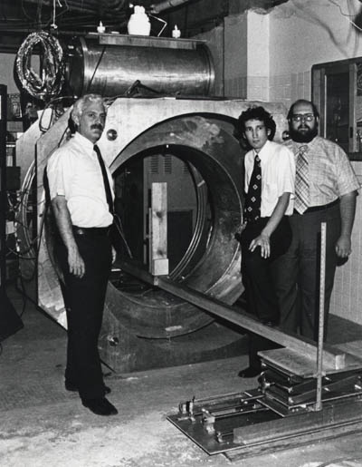
1977/1978 – Raymond Damadian went on to build the first MRI scanner by hand, assisted by his two post-doctoral students, Michael Goldsmith and Larry Minkoff at New York’s Downstate Medical Center and achieved the first MRI scan of a healthy human body in 1977 and a human body with cancer in 1978. Peter Mansfield improves mathematics behind MRI and develops echo-planar technique, which allows images to be produced in seconds and later becomes the basis for fast MR imaging.
1983 – Ljunggren and Tweig introduce k-space.
1986 – Le Bihan publishes an article in Radiology, which describes diffusion-weighted imaging (DWI).
1987 – Real-time MR imaging of the heart is developed.
1991 – Filler and colleagues describe imaging of axonal transport of supermagnetic metal oxide particles, a technique, which later becomes important in imaging of neural tracts.
1993 – Functional MR imaging of the brain is introduced.
1994 – The first intraoperative MR unit developed by GE and Harvard is installed in the Brigham and Women’s Hospital in Boston.
1990’s – In addition to research centres and large hospitals, small remote hospitals and imaging centres begin to utilize MRI predominantly for neuroimaging and musculoskeletal imaging.
2000’s – Cardiac MRI, Body MRI, fetal imaging, functional MR imaging are further developed and become routine in many imaging centres. Research centres make significant strides forward in imaging cartilage on high field scanners. The number of free-standing MRI centres, most of which utilize low or moderate field MR scanners significantly increases.
See more at http://www.two-views.com/mri-imaging/history.html#sthash.2BaGRUWw.dpuf
First MRI
MRI scans are delicate machinery that needs to be handled with care and proper installation knowledge to install it.
If guidelines are not followed properly the output image will be distorted and of poor quality.
Homogeneous Magnet are required
Magnets are shimed to get a homogenous magnetic field that gives you precise values. Some professionals do not shim the magnet which results in poor image quality.
Calibration is required to produce good images
A good image can be produced if RF gradient power, linearity and eddy currents are calibrated properly.
InBrain Innovations has a team of highly efficient engineers who are experts in various machines and we are well equipped with Tools that are required for the installation of the machines as per the manufacturer’s protocol. We have a group of engineers specialised on various machines and we have Tools to do as per the manufacturer’s installation protocol. We assure services for all image quality issues.
We have the experience of MRI installation over 40 countries. We have tie-ups with the top supplier who supply, Best MRI Machine in India. Our top suppliers are:
Hitachi MRI India


